Call us now
08071794481

 Send Inquiry
Model of Animal Mitosis
MOQ : 10 Pieces
Model of Animal Mitosis Specification
- Usage
- For Laboratory, College and Hospital Purpose
- Product Type
- Mitosis Model
- Material
- Hard Plastic
- Feature
- Long Working Life
- Color
- Mix Colour
Model of Animal Mitosis Trade Information
- Minimum Order Quantity
- 10 Pieces
- Payment Terms
- Cash in Advance (CID), Cash Advance (CA)
- Supply Ability
- 500 Pieces Per Day
- Delivery Time
- 1 Week
- Packaging Details
- Carton Box
- Main Export Market(s)
- Eastern Europe, Middle East, Central America, South America, Western Europe, Asia, North America, Australia, Africa
- Main Domestic Market
- All India
About Model of Animal Mitosis
Showing mitosis stages in animalcell Made of hard plastic. This model
will help clarify and provide a perfect
visual comparison of these two key
cell processes. This model identies
the parts of the cell phases of mitosis
and the changes cells undergo
through the stages of mitosis.
Nucleus, centrioles, centrosome,
chromatin, chromosomes, spindle,
aster and other key structures are
easily identied. Mounted on Board with key card.
Durable Material for Extended Use
Crafted from hard plastic, the model is built to withstand continuous handling in laboratories and classrooms. Its robust construction ensures it maintains its shape and detail even during frequent demonstrations. This long working life makes it a reliable choice for institutions seeking durable teaching aids.
Clear Visualization of Mitosis Stages
The model employs a mix of colors to vividly represent each phase of animal mitosis, aiding in easier differentiation and understanding. Students and professionals can explore the cell division process step by step, which promotes active engagement and better retention of complex biological concepts.
Versatile Applications
This model is specifically designed for use in laboratories, colleges, and hospitals. Its educational advantages make it suitable for demonstrating mitosis to students, training medical staff, and supporting academic research. As it is produced in India and exported internationally, it serves a wide range of users.
FAQs of Model of Animal Mitosis:
Q: How does the Model of Animal Mitosis help in understanding cell division?
A: The model visually demonstrates each stage of mitosis, allowing users to clearly observe the sequence and structure of cell division. The use of different colors for each phase helps clarify the process, making complex biological concepts easier to grasp for students and professionals alike.Q: What benefits does the hard plastic material provide for this mitosis model?
A: Hard plastic ensures durability and a long working life, even under frequent use in laboratory and educational environments. This robust material also allows the model to retain detailed features essential for effective teaching and demonstration.Q: Where can the Model of Animal Mitosis be used most effectively?
A: This model is best suited for laboratories, colleges, and hospitals, where it can be utilized for educational demonstrations, student experiments, and training sessions for medical professionals.Q: When is it appropriate to use this model in an educational setting?
A: The model is ideal for use during biology classes, lab sessions, or workshops when teaching or demonstrating the process of animal mitosis. It can also be used in assessments or practical exams to enhance experiential learning.Q: What is the process for using the model in teaching?
A: Educators can use the model by displaying each mitosis phase in sequence, using the mixed colors to guide explanations. Students can interact with the model, identify different phases, and better understand the chronological progression of cell division.Q: How does the mix of colors on the model benefit users?
A: The use of multiple colors distinguishes each phase of mitosis, helping users visually separate the stages and understand the specific changes that occur during cell division. This color-coding simplifies the learning process.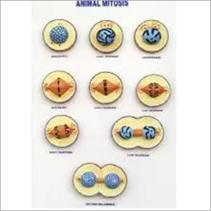
Tell us about your requirement

Price:
Quantity
Select Unit
- 50
- 100
- 200
- 250
- 500
- 1000+
Additional detail
Mobile number
Email

More Products in Biology & Earth Science Category
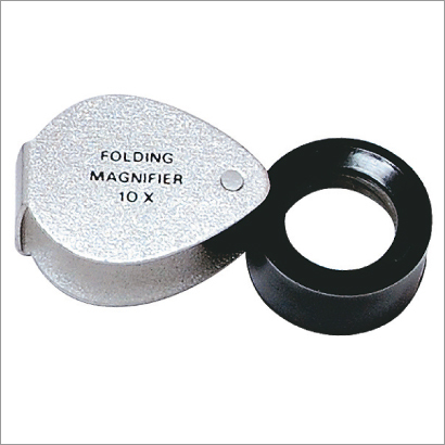
Magnifier Folding Pocket
Price Range 50.00 - 350.00 INR / Piece
Minimum Order Quantity : 10 Pieces
Material : Metal. Plastic and Glass
Feature : Durable and Better Efficiency
Usage : For Laboratory
Color : Silver and Black

Bone Shears Bone Cutting Forceps
Price Range 50.00 - 500.00 INR / Piece
Minimum Order Quantity : 10 Pieces
Material : Stainless Steel
Feature : High Performance
Usage : For Laboratory, College and Hospital
Color : Silver

Advance Student Microscope
Price Range 2000.00 - 10000.00 INR / Piece
Minimum Order Quantity : 10 Pieces
Material : Plastic, Metal and Glass
Feature : Durable and Better Efficiency
Usage : For Laboratory and RND
Color : White and Black
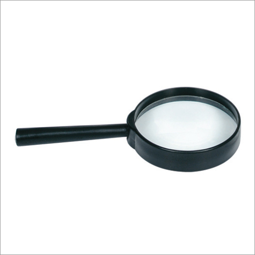
Plastic Magnifier
Price Range 50.00 - 450.00 INR / Piece
Minimum Order Quantity : 10 Pieces
Material : Plastic and Glass
Feature : Long Working Life and Better Efficiency
Usage : For Laboratory
Color : Black and Transparent
SADANA BROTHERS
SCI-LAB SOLUTION
SCI-LAB SOLUTION
Plot No- A-8/1, Naraina Ind. Area, Phase 1, New Delhi - 110028, India
Phone :08071794481
 Send Inquiry
 Send SMS
SADANA BROTHERS
All Rights Reserved.(Terms of Use)
Developed and Managed by Infocom Network Private Limited.
Developed and Managed by Infocom Network Private Limited.

