Call us now
08071794481

 Send Inquiry
Model Of Human Eye
MOQ : 10 Pieces
Model Of Human Eye Specification
- Usage
- For Laboratory, College and Hospital
- Product Type
- Eye Model
- Feature
- Durable and High Functionality
- Color
- White, Blue, Black and Red
Model Of Human Eye Trade Information
- Minimum Order Quantity
- 10 Pieces
- Payment Terms
- Cash in Advance (CID), Cash Advance (CA)
- Supply Ability
- 500 Pieces Per Day
- Delivery Time
- 1 Week
- Packaging Details
- Carton Box
- Main Export Market(s)
- Asia, Australia, North America, South America, Eastern Europe, Western Europe, Middle East, Central America, Africa
- Main Domestic Market
- All India
About Model Of Human Eye
Can be dismantled, showingall the basic artery, blood
vessel and other important
parts, Mounted on a stand
180 x 180 x 320mm. This
model is easy to take
apart and reassemble in
all aspects of internal
and external anatomy.
Removable parts include
the upper half of the sclera with cornea and muscle insertion. Two parts choroids with iris, lens and vitreous humor. Detail painting
highlights the iris, major blood vessels, and the origin of the optic
nerve. Mounted on base with key card.
Comprehensive Teaching Tool
This human eye model is engineered to facilitate clear, effective demonstrations in both academic and clinical environments. Its precise detailing enables users to visually explore the anatomical structure of the eye, benefiting students and educators alike. Ideal for lecture rooms, research labs, and hospital training, the model enhances understanding through interactive learning.
Durable and Functional Design
Built using high-quality materials, our model ensures lasting performance despite frequent use. The color combination of white, blue, black, and red depicts the natural anatomy faithfully, while its sturdy construction resists wear and tear. The models practical design supports in-depth study, frequent handling, and repeated assembling without loss of detail or integrity.
FAQs of Model Of Human Eye:
Q: How is the human eye model used in educational and healthcare settings?
A: The model is widely used for demonstrations, hands-on learning, and practical examinations in colleges, laboratories, and hospitals. It helps students, trainees, and professionals to visually analyze and understand the detailed structures and functions of the human eye.Q: What makes this eye model suitable for export and institutional use?
A: Its robust construction, accurate anatomical detailing, and high functionality make it ideal for exporters and institutions. The model is designed to withstand frequent handling and provides consistent educational value, meeting international standards for quality and durability.Q: When should institutions consider using this anatomical eye model?
A: Institutions should incorporate this model during anatomy lessons, practical laboratory sessions, staff training, and patient education initiatives. It is especially beneficial when a tangible, 3D representation of the human eye is required for better comprehension.Q: Where is this model of the human eye manufactured?
A: The human eye model is manufactured in India, delivered by experienced exporters and manufacturers with a strong reputation for supplying high-quality anatomical models to institutions worldwide.Q: What is the benefit of the multicolored design in the eye model?
A: The use of white, blue, black, and red colors highlights different anatomical regions, aiding clearer differentiation and enhanced visualization. This feature supports teaching by allowing users to easily identify key structures and relationships within the eye.Q: What process is followed to ensure the models durability and functionality?
A: The model is crafted from durable materials and undergoes quality assurance tests for strength, color retention, and anatomical accuracy. This process ensures that the product maintains its high functionality throughout extended periods of educational use.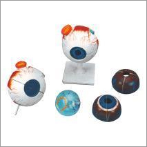
Tell us about your requirement

Price:
Quantity
Select Unit
- 50
- 100
- 200
- 250
- 500
- 1000+
Additional detail
Mobile number
Email

More Products in Biology & Earth Science Category

Lab Tulgreen Funnel
Price Range 1000.00 - 5000.00 INR / Piece
Minimum Order Quantity : 10 Pieces
Color : Black and White
Product Type : Tulgreen Funnel
Usage : For Collecting Organisms From Soil
Feature : Durable and Better Efficiency

Glass Culture Bottle
Price Range 50.00 - 200.00 INR / Piece
Minimum Order Quantity : 10 Pieces
Color : Transparent
Product Type : Culture Bottle
Usage : For Laboratory, College and Hospital Purpose
Feature : Long Working Life

White Plastic and Glass Photosynthesis Apparatus
Price Range 250.00 - 1500.00 INR / Piece
Minimum Order Quantity : 10 Pieces
Color : White and Black
Product Type : Photosynthesis Apparatus
Usage : For Measurement and Analysis of gas evolved by water plants under Different Light Conditions
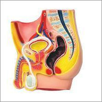
Model Of Human Male Pelvis
Price Range 2000.00 - 6000.00 INR / Piece
Minimum Order Quantity : 10 Pieces
Color : Red and Yellow
Product Type : Male Pelvis Model
Usage : For Laboratory and RND
Feature : Long Working Life and High Functionality
SADANA BROTHERS
SCI-LAB SOLUTION
SCI-LAB SOLUTION
Plot No- A-8/1, Naraina Ind. Area, Phase 1, New Delhi - 110028, India
Phone :08071794481
 Send Inquiry
 Send SMS
SADANA BROTHERS
All Rights Reserved.(Terms of Use)
Developed and Managed by Infocom Network Private Limited.
Developed and Managed by Infocom Network Private Limited.

