Call us now
08071794481

 Send Inquiry
Model Of Human Ear
MOQ : 10 Pieces
Model Of Human Ear Specification
- Usage
- For Laboratory, College and Hospital
- Product Type
- Ear Model
- Feature
- Durable and High Functionality
- Color
- White, Yellow, Red and Pick
Model Of Human Ear Trade Information
- Minimum Order Quantity
- 10 Pieces
- Payment Terms
- Cash in Advance (CID), Cash Advance (CA)
- Supply Ability
- 500 Pieces Per Day
- Delivery Time
- 1 Week
- Packaging Details
- Carton Box
- Main Export Market(s)
- Western Europe, Australia, South America, Middle East, Central America, Asia, Eastern Europe, North America, Africa
- Main Domestic Market
- All India
About Model Of Human Ear
This model shows pinnawith auditory canal,
tympanic membrane,
Eustachian tube, middle
ear ossicles, bony labyrinth
and cochlea. Tympanic
membrane, middle and
inner ear. Removable
part include the tympanic
membrane (eardrum) with malleus and incus (hammer and anvil),
and the two part labyrinth / cochlea with stapes (stirrup) and
auditory nerve. Enlarged 3 times. Includes mounting /display base
and key card.
Accurate Educational Aid
This human ear model vividly illustrates anatomical details in realistic colors, supporting medical students, faculty, and healthcare professionals in learning and teaching activities. Its accuracy makes it a dependable resource for lectures, practical sessions, and patient education.
Built to Last
Designed with durability in mind, the model withstands frequent classroom or laboratory use. Its sturdy construction ensures function over many semesters, making it a cost-effective investment for institutions.
FAQs of Model Of Human Ear:
Q: How is the Model of Human Ear used in laboratories and hospitals?
A: The model is employed in laboratories and hospitals to visually demonstrate the structure and function of the human ear, facilitating practical learning for students and aiding medical professionals in explaining ear anatomy to patients.Q: What features make the Human Ear Model highly functional for educational purposes?
A: This models detailed representation, combined with color-coded anatomical sections, enables effective visualization and understanding of ear parts. Its durability allows repeated use without compromising quality, making it especially suitable for educational settings.Q: When should institutions consider investing in this Human Ear Model?
A: Institutions should consider acquiring this model when they require a reliable and long-lasting teaching aid for anatomy classes, medical demonstrations, or public health education, especially where repeated handling is expected.Q: Where can the Model of Human Ear be used effectively?
A: It is most effectively used in colleges, medical laboratories, and hospitals, where detailed anatomical education or demonstrations are needed for students, healthcare trainees, or patient awareness initiatives.Q: What is the manufacturing process behind this ear model?
A: The manufacturing process involves skilled craftsmanship using high-quality materials to ensure durability and precise anatomical detailing. Each model is carefully colored in white, yellow, red, and pink to enhance the visualization of different ear components.Q: What are the benefits of using this model in educational settings?
A: Using this model allows for hands-on learning, improved comprehension of ear anatomy, and enables educators to demonstrate concepts interactively. Its robust design supports frequent usage, making it a practical choice for busy learning environments.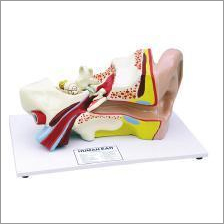
Tell us about your requirement

Price:
Quantity
Select Unit
- 50
- 100
- 200
- 250
- 500
- 1000+
Additional detail
Mobile number
Email

More Products in Biology & Earth Science Category
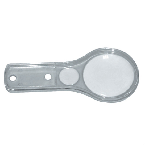
Clear Plastic Magnifier
Price Range 52.00 - 380.00 INR / Piece
Minimum Order Quantity : 10 Pieces
Feature : Long Working Life and Better Efficiency
Usage : For Laboratory
Product Type : Plastic Magnifier
Color : Clear
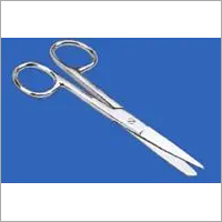
Scissors Dissecting Straight
Price Range 120.00 - 750.00 INR / Piece
Minimum Order Quantity : 10 Pieces
Feature : Long Working Life and High Strength
Usage : For Fine Dissection Work
Product Type : IRIS Curved Scissors
Color : Silver
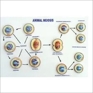
Model of Meiosis
Price Range 130.00 - 1000.00 INR / Piece
Minimum Order Quantity : 10 Pieces
Feature : Long Working Life
Usage : For Laboratory, College and Hospital Purpose
Product Type : Meiosis Model
Color : Mix Colour
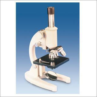
Student Microscope (Senior)
Price Range 1500.00 - 5000.00 INR / Piece
Minimum Order Quantity : 10 Pieces
Feature : Durable and Better Efficiency
Usage : For Laboratory and RND
Product Type : Microscope
Color : White and Black
SADANA BROTHERS
SCI-LAB SOLUTION
SCI-LAB SOLUTION
Plot No- A-8/1, Naraina Ind. Area, Phase 1, New Delhi - 110028, India
Phone :08071794481
 Send Inquiry
 Send SMS
SADANA BROTHERS
All Rights Reserved.(Terms of Use)
Developed and Managed by Infocom Network Private Limited.
Developed and Managed by Infocom Network Private Limited.

